Wednesday, December 12, 2012

Microscope - Student Monocular Biological Microscope 40x-400x with Light

Microscope - Student Monocular Biological Microscope 40x-400x with Light Review


Prices as of : Dec 12, 2012 02:00:03
Listprice :
Offier Price :
You Save :

Microscope - Student Monocular Biological Microscope 40x-400x with Light Feature

  • Monocular head included at 45 degrees, 10X zoom wide focus eyepiece, objective lens at 4X, 10X, and 40X for 40x, 100x & 400x magnifications
  • Plain 90mm x 90mm stage with clips
  • Single lens condenser with Disc diaphragm
  • Coarse adjustment focusing
  • Powered by 110/20W Incandescent lamp, requires available standard electrical outlet

Microscope - Student Monocular Biological Microscope 40x-400x with Light Overview

This microscope is of metal construction and built to last. Magnify objects from 40 to 400 times with three objectives - 4x, 10x, and 40x, and a widefield 10x eyepiece. The stage measures 90mm x 90mm with two spring clips to retain the slides. This microscope is a perfect solution for schools and families looking to introduce the concepts of microscopy to young students without the expense of a more advanced instrument. This microscope stands 292mm high. The base measures 152mm x 114mm. Specifications: Monocular head included at 45 degrees, 10X zoom wide focus eyepiece, objective lens at 4X, 10X, and 40X. Plain 90mm x 90mm stage with clips, single lens condenser with Disc diaphragm, coarse adjustment focusing. Powered by 110/20W Incandescent lamp. Requires available standard electrical outlet.

*** Product Information and Prices Stored: Dec 12, 2012 02:00:03

Thanks To : Pneumatic Plumbing Picking Safety Products Finishing Products Sonic Screwdriver Tool

Monday, December 10, 2012

Celestron 44302 Deluxe Handheld Digital Microscope 2MP

Celestron 44302 Deluxe Handheld Digital Microscope 2MP Review


Prices as of : Dec 10, 2012 14:00:04
Listprice : $81.95
Offier Price : $39.99
You Save : $41.96

Celestron 44302 Deluxe Handheld Digital Microscope 2MP Feature

  • Handheld Digital Microscope Powered by a 2.0 USB Cable.Digital Camera - built-in 2MP for Snapshot Images and Videos
  • Computer Requirements - UVC Plug and Play with Windows 7, Vista, and XP and MAC 10.4.9 and later, CD/DVD Drive and open USB Port
  • Power - 10x to 40x and 150x
  • LED Illumination.
  • CD-ROM - Software for Basic Image Capture and Organization as well as Simple Measurement Function (on Windows based operating systems)

Celestron 44302 Deluxe Handheld Digital Microscope 2MP Overview

This innovative microscope design allows you to view specimens or objects on a computer screen and allows you to take snapshot images or short videos. A rugged, metal stand is included for holding the microscope steady at various positions for viewing and/or imaging.

*** Product Information and Prices Stored: Dec 10, 2012 14:00:04

Related : Pneumatic Plumbing Best Fasteners Sun Solar cell kodak camera easy share

Sunday, December 9, 2012

Best Microscopes For Kids - beautiful Tips to make your mind up the Best Microscope

Microscope is a breathtaking scientific invention that made it potential to study finer details that are present in the structure. This will also make it potential to learn about assorted kinds of microorganisms that are present in the environment. Microscopes are being widely used in assorted kinds of fields such as medical, physics, chemistry and many more in a detailed manner.

There are assorted kinds of these devices that are available in the market. There has been breathtaking amelioration that has taken place in the field of lens making and thus there is great opening that you will be able to get breathtaking microscopes at thrifty costs. Microscope is essentially a blend of assorted kinds of lens that will make it potential to study about assorted objects. Some of the beloved microscopes that are available include dissection and stereo.

Light Source Microscope

The two leading features that should be looked upon while buying the best microscopes for kids are in case,granted below.

Light source

The light source for microscope most ordinarily involves a mirror or a small bulb. The whole of light that is being focused on the object should be of optimum level. There should not bee too low or too appealing images that will not be providing permissible images. You shall potential stay way from cheap microscopes that will be using mirrors as a light source. A lot of kids love what is being in case,granted by small light bulbs to visualize assorted features around them

The ratio is great

Another leading aspect that should be looked upon while purchasing these microscopes is the magnification ratio. It will be good to buy devices that will be providing magnification of the order of 10x to 40x. Many habitancy are greatly satisfied with these magnification factor to visualize assorted kinds of features such as flowers, insects and many more that is present in day to day life. The cost of these microscopes will also be of affordable level.

Best Microscopes For Kids - beautiful Tips to make your mind up the Best Microscope

Visit : Best Fasteners The Fasteners signal booster for cell phone Server Rack Rails

Saturday, December 8, 2012

Types of combination visual Microscopes

Compound optical microscopes are also known as combination microscopes, light microscopes or optical microscopes and come in some varieties. These comprise digital, inverted, stereo, monocular and binocular.

All work on the same system and deliver an enlarged image to the viewer. They have some differences though, which will be detailed here.

Microscope

Monocular:

These are probably the ones you think of when microscopes come to mind. Using a singular light as a sample illuminator and combination lenses for magnification, these have a singular eyepiece. This is a bit uncomfortable, as you need to close one eye to get a clear image of what you are looking at on the slide. The eyepiece has a power of about 10X and the objectives or lenses on the nosepiece range from 2X to 50X depending on your singular microscope. The big one here is the singular eyepiece.

Binocular:

Binocular microscopes are becoming more common. You can tell a binocular style microscope by the dual eyepiece. Plainly put, you use both eyes to view the sample image. This makes them more comfortable to use and thereby more popular. Dual eyepiece scopes are used widely in high school and college classrooms. Binocular microscopes have all the same characteristics as the monocular ones above.

Stereo:

Stereo microscopes bring a whole new size to the image, literally. Whereas the typical light microscope produces a two-dimensional image, the stereo microscope uses two light sources working independently to produce a three-dimensional image to the viewer. The sample on the slide will have height, depth and width. Using all of the same features and characteristics of the other optical microscopes, stereomicroscopes stand out among their counterparts.

Digital:

This is where things go differently in the realms of microscopy. Approved optical microscopes use light to illuminate and lenses to magnify something that you could not see with the unaided eye.

Digital microscopes are a breed apart. This type of microscope inverts the light source, placing it above the sample. Approved optical microscopes place the light below the sample. You also get the 3D image like in stereo scopes. The inequity is that the image is digitized and transmitted to a monitor or screen for viewing. Imagine watching cells divide on a 19" monitor. The user can take still photos or thoughprovoking video of the sample in real time. I think you can see the advantages here.

Inverted:

Inverted microscopes are used to study samples that are gravity sensitive, like gases suspended in a liquid. The inversion refers to the light source, which is typically below the sample slide. With the inverted microscope, the light source is above the sample slide. This paved the way for new innovations in digital imaging that came along.

All these dissimilar types will work for you. It's just a matter of what you want to perform or study. I can tell you this, though; using any microscope will open your eyes to a whole new perspective of where we came from and what we are made of.

Types of combination visual Microscopes

My Links : Best Fasteners Finishing Products Server Rack Rails solar power cell phone charger

Friday, December 7, 2012

History And Uses Of optic Microscopes

Optical microscopes use visible light and a ideas of lenses to magnify small samples that are usually un-seen to the bare eye. The optic microscope is the first, oldest and simples type of microscope (as opposed to the much more advanced electronic microscope). The first optic microscopes were created in the 18th century. Due to it's contract sizes, simplicity and relatively low price, the optic microscope is very popular, and can be found in use in many areas of biology. optic microscopes mostly magnify objects for up to 1500 times.

The first optic microscopes were structured in a way that is called "the simple microscope". This buildings utilizes only one pair of lenses to create a magnified image of the sample. Today, the simple buildings is in use only in the magnifying glass, hand lens and the loupe.

Microscope

The more advances optic microscopes, and the ones that are favorite today, are what's called "compound optic microscopes". These microscopes use a ideas of many lenses, in order to "compound" and multiply the magnification, and therefore maximize it. The two main lens systems in an optic microscope are the objective lens (near the examined object), and the eyepiece lens (up near the eye of the scientist). Modern optic microscopes use many lenses both in the objective part as well as the eyepiece part.
The old optic microscopes also used a mirror to furnish illumination below the object. The Modern optic microscopes use a strong lamp to furnish constant and strong illumination.

So what are optic microscopes used for now a days?
The main uses of compound optic microscopes include:

The examining small pieces of material, or even a smear or a squash preparation. This is due to the fact that the optic microscope uses light to pass beneath the object and enter the lenses. That's why the item is best be half-transparent. In other uses the optic microscope may be used to gawk metal samples, in order to study the metal's structure.
At low power, microscopes can be used to gawk small living animals and plants. At high power, they can be used to gawk bacteria.

It is foremost to note that the vast advancement in remedial fields and biology in general, is owed to a large extent, to the invention of the optic microscopes. For example, the way the blood flows in our body was not fully understood until the microscope made in inherent to gawk small blood vessels behavior.

History And Uses Of optic Microscopes

Recommend : Pneumatics and Plumbing Good choice high definition media player Case Rackmount

Thursday, December 6, 2012

Student Microscope Set

Student Microscope Set Review


Prices as of : Dec 06, 2012 04:30:03
Listprice :
Offier Price :
You Save :

Student Microscope Set Overview

Student Microscope Set. This science explorer microscope set from Magic Cabin helps develop an early interest in science, and encourages children to learn more about nature and the world around them. The 62-piece set includes everything needed to look at small living objects, including the high-quality microscope itself, slides, a Petri dish, pipettes, a stirring rod and even a screwdriver and spare bulb! The microscope features magnification to 100x, 450x and 900x with changeable lenses, so a child can get up close and personal with many different objects and miniscule life forms. Ages 8 and up. 62-Piece Deluxe Student Microscope Set develops an interest in science or nature, and encourages children to learn about their world Microscope is made of die-cast metal to withstand heavy use and includes three lenses to magnify objects to 100x, 450x and 900x Set includes 62 pieces, from slides, pipettes, a Petri dish and vials to labels, a screwdriver, a spare bulb and a Micro-science databank card 4-3/4L x 3W x 8-1/2 microscope and included pieces come in a carrying case to keep everything organized and easy to move This science explorer microscope set was created for children aged 8 and older Size Microscope: approx. 4-3/4"L x 3"W x 8-1/2"H

*** Product Information and Prices Stored: Dec 06, 2012 04:30:03

Visit : The Pneumatic Fasteners Equipment Finishing Products cell phone covers samsung phones hd blu ray player

Tuesday, December 4, 2012

AmScope 10x-20x-30x-60x Digital Binocular Stereo Microscope with Top & Bottom Dual Light Illumination System + USB Digital Camera

AmScope 10x-20x-30x-60x Digital Binocular Stereo Microscope with Top & Bottom Dual Light Illumination System + USB Digital Camera Review


Prices as of : Dec 05, 2012 00:15:03
Listprice : $800.00
Offier Price : $179.98
You Save : $620.02

AmScope 10x-20x-30x-60x Digital Binocular Stereo Microscope with Top & Bottom Dual Light Illumination System + USB Digital Camera Feature

  • Four Magnification Power Settings: 10X, 20X, 30X & 60X
  • Built-In Both Incident (top) Lighting and Transmitted (bottom) Lighting
  • Dispays real-time live images with 640x480 pixel high resolution
  • Captures microscope still images, records live videos, and saves images in BMP, TIFF, JPG, PICT, PTL and other formats
  • Advanced editing, processing measuring software compatible with Windows 2000/XP/Vista/7

AmScope 10x-20x-30x-60x Digital Binocular Stereo Microscope with Top & Bottom Dual Light Illumination System + USB Digital Camera Overview

This binocular arm-stand stereo microscope comes with a 640x480 pixel USB digital camera. It has four magnification power settings, 10X, 20X, 30X & 60X, and a versatile illumination system that provides both incident (top) lighting and transmitted (bottom) lighting. You can choose between incident illumination shining down onto the object or transmitted illumination through the frosted stage plate. The first is used for the observation of three-dimensional objects and the second for the observation of slides. This microscope offers high resolution and good depth within a broad field of view. It gives sharp clear stereo images. Its 45° inclined binocular head ensures an easy observation and rubber eyeguards provide further comfort. The digital camera captures microscope images and displays live video on your computer screen. With the user-friendly software included, compatible with Windows 2000/XP/Vista/7, you can edit microscope images, record video, capture still images and save microscope images as a BMP, TIFF, JPG, PICT, PTL or other file. The camera dose not need any driver and the software is very easy to be installed on your computer. This microscopy system is a great instrument for many applications. It can be used in geology, forestry, archaeology, agriculture and biology. It is appropriate for educational purposes in hospitals and schools. It is great for the exploration of the world of surface details and fine structures of various materials like wood, metal, solder, semiconductor and stone. It is perfect for the examination of insects, plants, rocks, stamps, coins, jewel, soldered points, microelectronic components or virtually any three dimensional objects. This scope is an ideal tool for watchmaker, jeweler, engraver, dies-maker, philatelist, coin collector, students, and etc. It comes with first-class quality and affordable price. It is brand new in factory-sealed box. The retail value of this microscope is 0.

*** Product Information and Prices Stored: Dec 05, 2012 00:15:03

Thanks To : Best Fasteners Material Handing Products The Pneumatic canon easy share camera

Monday, December 3, 2012

Don't Confuse Your Baby's Yeast Infections With Diaper Rash

Yeast infections that affect a baby's diaper area not the same thing as diaper rash. Unfortunately, many well meaning parents use regular diaper cream and baby powder on the yeast infection and this will do nothing to cure it.

A yeast infection is dissimilar from a diaper rash in that it appears as a flat, red rash with scalloped edges that have some tiny rashes colse to it. A yeast infection rash may increase along the pubic area and onto the lower part of the baby's abdomen.

Light Source Microscope

The yeast infections that affect babies are called Candidas albicans, which is the most tasteless cause of all types of yeast infections. A pediatrician can decree whether a rash on the groin or a thrush in the mouth of babies is a yeast infection. To get a specific pathology the doctor may survey the scrapings of a yeast infection under a microscope.

Babies who are on antibiotics or who are being breastfed by mothers who are taking antibiotics are at higher risk for yeast infections. That's because antibiotics kill the good bacteria in the body that keep yeast in check. Without these bacteria around, yeast and grow more abundantly.

While applying diaper cream may bring your baby temporary relief from the itching and soreness it will not heal the infection. Without allowable treatment, a yeast infection in the baby's diaper area will get progressively worse.

Anti-fungal medications are the only types of medicine that can get rid of yeast infections. Some pediatricians might write a prescription for Nystatin while others may suggest and over the counter medicine like Lotrim Af to treat the diaper yeast infection.

Using antifungal cream, soaking the baby in warm water two times a day and frequent diaper changes will normally clear up a yeast infection within a few days. While you are treating the yeast infection make sure that your baby's diapers are loose enough to let air in. When changing your child's diapers, make sure the skin on his buttocks and groin area is clean and dry.

Some holistic medical practitioners advocate the use of a diaper cream containing tea tree oil to help clear yeast infections in the diaper area. Others suggest using aloe or pressing damp chamomile teabags directly on the baby's rash. If the yeast persists for more than a week or if your baby has fever or chills you should consult with your pediatrician.

Yeast infections in baby's can occur despite our best efforts. However, frequent diaper changes can decrease the chances of an infection occurring. Children with yeast infections in the diaper area should not be excluded from daycare because these types of infections are not contagious.

If your baby recently had thrush (a yeast infection in the mouth), her or she may end up with a yeast infection in the diaper area, too. That's because yeast passes straight through your baby's digestive principles when he eats and winds up in his stool, which ultimately lands in his diaper right next to his warm, damp skin. Yeast thrives in warm, damp environments.

Don't Confuse Your Baby's Yeast Infections With Diaper Rash

Tags : Picking Safety Products led microscope light media player hard disk

Saturday, December 1, 2012

12 Creative Photography Ideas

Here are 12 ideas to get your creative juices flowing and growth your skills and value as a photographer.

Tip 1 - Time Lapse photos. I showed my niece how to do this with clay figures and stitch the resulting frames together into a video file, and she was busy for days. If you control the exposure consistently to keep the images consistent with one another, you can do some beyond doubt fun stop-motion animation. Or, you can set up your camera to capture other slow motion effects such as flowers chance and seedlings growing.

Light Source Microscope

Tip 2 - Night Lights. Things look very dissimilar at night. Shooting city scenes with ready light creates some enchanting images. And shooting outdoor images under moonlight or with "light painting", where you open up the camera shutter for an extended exposure, and "paint" your targets with colored or plain light, can create some truly bizarre images.

Tip 3 - Astrophotography. Hook that Slr up to a telescope, and you are ready to peer into the depths of space and time. You'll need some adapters, and capability to compensate for the earth's rotation for beyond doubt long shots. Start with the moon and move on from there.

Tip 4 - Macro photography. From flowers to coins to stamps, you can polish your skills at close-up photography and capture some beyond doubt detailed images. Often a macro lens or close-up attachment will help. See my tips on Flower Photography to get more information.

Tip 5 - Micro photography. If you can interface that camera with a microscope, you can get some beyond doubt crazy images. Or, stack up a bunch of close-up magnification and try your hand at turning salt crystals into surreal imagery.

Tip 6 - guarnatee Photos. Ok, maybe a bit boring, but you and your friends and relatives will thank you. Take a merge hours and touch and photograph all things of value, with a full shot or two if each item of value, accompanied by a shot of the identifying marks - manufacturer model or serial number. Then burn a Cd or Dvd and store it off site. If you have a fire or other loss, this could save the owner thousands of dollars.

Tip 7 - house formula book. Anytime those house favorites are prepared, copy down the formula and take some photos of the food. You can yield a printed or electronic cookbook of house favorites that every person will love.

Tip 8 - Stock Photography. This is a very busy market niche, but the cost of entry is low. Specialize in things you love, and you may be able to create some income from your stock images. Hunt for stock photography sites, and make sure you understand your possession before you post images.

Tip 9 - special Effects. Maybe you want to specialize in high-speed images of athletes, or surrealistic collages. Try your hand at using your editing skills to put person in a soda bottle or floating on a candy lifesaver. Often more artistic than photographic, it will test your composition, lighting and editing skills to come up with believable artificial realities.

Tip 10 - Still Life. Ahh, the bowl of fruit. Sometimes a simple object or collection, properly lit, shot and edited, is a thing of beauty. It's a great way to study light. Start with an egg on a light background, a lamp and a window and see how you can learn about lighting and composition.

Tip 11 - Computer Control. Many cameras have a Usb interface and remote control software. You can beyond doubt control the camera from the computer. See if you can get it to work to your liking, and maybe even program some time lapse or exposure bracketing experiments.

Tip 12 - Be Like Andy. Take some images of everyday items and try to create those neat colored backgrounds like Andy Warhol used to make. create a 4-up print of the same image and change the colors of each quadrant to make an enchanting quadrilateral print.

Have fun with these ideas, and let me know when you come to be famous!

12 Creative Photography Ideas

Visit : Fasteners Equipment light microscope image easy share digital camera

Friday, November 30, 2012

AmScope 144 LED Adjustable Microscope Compact Ring Light + Adapter

AmScope 144 LED Adjustable Microscope Compact Ring Light + Adapter Review


Prices as of : Nov 30, 2012 16:30:03
Listprice : $320.00
Offier Price : $64.98
You Save : $255.02

AmScope 144 LED Adjustable Microscope Compact Ring Light + Adapter Feature

  • 144-LED ring light with compact design and adjustable brightness
  • Provides intense and focused shadow-free illumination
  • 2.4" (61mm) inside and 4" (100mm) overall outside diameters
  • Power input: 110-240V, 50-60HZ, auto switching
  • 1-7/8" (48 mm in diameter) ring adapter included

AmScope 144 LED Adjustable Microscope Compact Ring Light + Adapter Overview

This is a 144-LED intensity adjustable ring light for stereo microscopes with mounting size 2.4" (61mm) in diameter. The ring light has 2.4" (61mm) inside and 4" (100mm) overall outside diameters. It works with power supplies from a 100-240V and comes with a 1-7/8" (48 mm in diameter) ring adapter. With variable intensity control, it provides cool, even, intense and focused shadow-free illumination. It is an ideal durable cool light source for all stereo microscopes. This kit comes with one year warranty against manufacturing defects. It is brand new in original box. Its retail value is 0.

*** Product Information and Prices Stored: Nov 30, 2012 16:30:03

See Also : Measurement Guide Top 10 Tablet PCs

Thursday, November 29, 2012

Vivitar 20-Piece Microscope and Telescope Combo Bundle

Vivitar 20-Piece Microscope and Telescope Combo Bundle Review


Prices as of : Nov 29, 2012 09:00:05
Listprice : $59.99
Offier Price :
You Save :

Vivitar 20-Piece Microscope and Telescope Combo Bundle Feature

  • Includes 60x/120x Refractor Power Telescope & 900x Power Microscope
  • Lightweight Durable Construction
  • Quick Screw-Type Assembly
  • The Telescope Is Great For Star Tracking
  • The Microscope Is Great For Finding The Smallest Molecules Under The Eye

Vivitar 20-Piece Microscope and Telescope Combo Bundle Overview

This is a great beginner level biological microscope with advanced features. The electric illuminator provides direct control of light. A mechanical stage is standard equipment with knobs controlling both axes for easy centering of specimen slides. This Vivitar microscope set is a 20 piece set which includes: A microscope with 3 Adjustable lenses and a Illuminator lamp, 3 plastic vials and 3 plastic slide covers, a stirring rod, spatula, scalpel, and needle pen, 3 collecting stickers, and 2 circular specimen trays.

*** Product Information and Prices Stored: Nov 29, 2012 09:00:05

Friends Link : Measurement Guide blu ray player hd wifi waterproof digital camera canon

Wednesday, November 28, 2012

insight the Power of a combination Microscope

Probably a tasteless apparatus at most high school science laboratories, the mixture microscope use the beloved mixture of dual lenses and light reflection. In a world becoming infinitesimally smaller in each generation, microscopes of all types, caliber, sizes and capabilities can be found virtually everywhere - from laboratories in high school campuses, colleges, universities, to study and improvement laboratories of pharmaceutical associates and a host of other manufacturers.

Getting to Know Microscopes

Light Source Microscope

The mixture microscope has more than one lens - commonly two - to do its job. The most basic of all types - the uncomplicated microscope uses a single lens. Impliedly, it offers a good view and more capabilities. Here are basics of microscopy:

1. Light Source.

Illumination is a basic requirement in microscopy. Some microscopes have mirrors that reflect light from sources exterior of the microscope. Others, like the fluorescence microscope have their own definite sources of illumination, in its case a mercury-vapor lamp or a xenon arc lamp. Those using definite light sources are ordinarily more mighty as these lights are more intense than generic day or lamplights used by ordinary mixture microscopes.

2. Objective Lens.

All microscopes have objective lens - the lens closest to the object under probe; not all microscopes have eyepieces. Although the tasteless image the word conjures is that of an ocular device, there are extremely industrialized microscopes that do not have an eyepiece. Some microscopes have three eyepieces for normal viewing and for mounting a camera, like the trinocular microscope. Instead, there are mounted on cameras and or video displays for a state-of-the-art, 3D view of minute objects or organisms. If you follow beloved American Tv, you would have already seen this in shows like House, Md.

3. Magnification Controls.

Yes, the term sounds impressive but it's also very basic. Microscopes, regardless of power and/or grade, allow you to operate magnification levels with adjustment knobs. Usually, the magnification power is a factor of the objective lens and the eyepiece. In all cases, the maximum magnification is 2000x.

Practical Applications

Compound microscopes are used in many fields and for various purposes. The type of mixture microscope you need and the magnification levels you wish is determined by what you want to see, the specimen you're using and what you want to do with the images. Incidentally, manufacturers have industrialized a range of microscopes with definite applications and users in mind. It's now tasteless to find dissimilar grades of one type of microscope. Student-grade mixture microscopes - probably an inexpensive tool - are found in virtually all high school laboratories over the globe. Professional-grade mixture microscopes can be found in study laboratories where you commonly find white coats. Fortunately, manufacturers of this equipment like Meji Techno, Nikon, and Olympus make customizing microscopes potential with a range of selections that come with various accessories. You can configure your own microscope to suit your definite application.

But wait! That's not all. Working closely with its user base, these associates are permanently working toward improving their existing line with creative and imaginative input from population who use their products the most. So, have you and your mixture microscope met?

insight the Power of a combination Microscope

Friends Link : Finishing Products The Fasteners Xtreme Titanium Watch Moblie Mount

Tuesday, November 27, 2012

Parts and Functions of a Light Microscope (Part Ii)

A primitive microscope was invented in 1590 in Middelburg, Netherlands, by the eyeglass makers Hans Lippershey, Zacharias Jansen and his father Hans Jansen. Further, Galileo Galilei improved the instrument by using a set of aligned lenses and called it "occhiolino", what means "little eye". In 1625, Giovanni Faber named Galileo Galilei's "occhiolino" as a mixture microscope and this name remains until today.

The optic microscope, the most common type of microscope, contains several parts with exact functions. examine the picture and find their functions.

Microscope

1. Eyepiece: contains the ocular lens, which provides a magnification power of 10x to 15x, usually. This is where you look through.

2. Nosepiece: holds the objective lenses and can be rotated no ifs ands or buts to turn magnification.

3. Objective lenses: usually, there are three or four objective lenses on a microscope, consisting of 4x, 10x, 40x and 100x magnification powers. In order to derive the total magnification of an image, you need to multiply the eyepiece lens power by the objective lens power. So, if you couple a 10x eyepiece lens with a 40x objective lens, the total magnification is of 10 x 40 = 400 times.

4. Stage clips: hold the slide in place.

5. Stage: it is a flat platform that supports the slide being analyzed.

6. Diaphragm: it controls the intensity and size of the cone light projected on the specimen. As a rule of thumb, the more transparent the specimen, less light is required.

7. Light source: it projects light upwards straight through the diaphragm, slide and lenses.

8. Base: supports the microscope.

9. Condenser lens: it helps to focus the light onto the sample analyzed. They are particularly helpful when coupled with the top objective lens.

10. Arm: supports the microscope when carried.

11. common adjustment knob: when the knob is turned, the stage moves up or down, in order to common adjust the focus.

12. Fine adjustment knob: used fine adjust the focus.

Visit my blog for the next article: ideas and application of light microscopy.

Parts and Functions of a Light Microscope (Part Ii)

Recommend : Fasteners Equipment The Pneumatic cell phone plans for kids microscope lights

Sunday, November 25, 2012

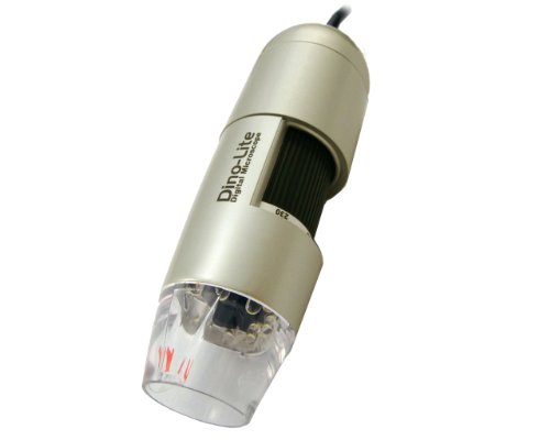
Dino-Lite Digital Microscope (USB2.0) 10x~200x Magnification

Dino-Lite Digital Microscope (USB2.0) 10x~200x Magnification Review


Prices as of : Nov 25, 2012 18:30:05
Listprice : $200.00
Offier Price : $149.00
You Save : $51

Dino-Lite Digital Microscope (USB2.0) 10x~200x Magnification Feature

  • ** Stand shown in picture is sold seperately!
  • Output: PC/USB, Magnification: 10x-200x, illumination: 8 LED
  • Image Capture: JPEG/BMP 640x480 Video Capture: AVI 640x480 30fps
  • Enabling you to capture image, video and time-lapsed video in a click
  • Contents: Camera, manual, Dino Capture software compatible w/ all Windows and Mac OS 9, X

Dino-Lite Digital Microscope (USB2.0) 10x~200x Magnification Overview

** Stand shown in the picture is sold seperately. Dino-Lite Digital Microscope is an amazing and easy to use handheld microscope. It is ideal for various applications such as skin check, scalp check, science learning, visual assistance and industrial inspections. Features include magnification up to 200 times(see model comparison chart below), capability of interfacing to PC, adjustable focus, quick and easy installation, and stylish design.Dino digital microscope provides high quality microscopy video interfacing to PC. Clear and steady imaging with simple magnification and focus control.The bundled software, "DinoCapture", allows the user to take a snapshot, video or time-lapsed video with just a click on TV or Computer. A user friendly interface to create a movie in AVI format. The video quality can be adjusted by video compression setting. Multiple pictures can be easily compared on screen as well! The range of magnifications goes from 10X to 200X for great selection in image viewing. The white LED light equipped with the digital microscope will enhance the illumination to improve resolution.Images and Data can be stored in the computer hard drive, CD, DVD or Polaroid and Video.The emailing function included with the software allows for the instant sending of images from remote locations around the world.

*** Product Information and Prices Stored: Nov 25, 2012 18:30:05

Related : Pneumatic Plumbing Best Fasteners Rackmount 19 inch Choosing The Correct Tripods

Saturday, November 24, 2012

GSI High-Definition Scientific Digital LED Microscope, USB Video Connection to Computers/Laptops/Notebooks, Magnifies All Microscopic Articles 200x, Special Image Treatment, Includes Stand for Handsfree Use, for Hobby/Science/Education/Industrial

GSI High-Definition Scientific Digital LED Microscope, USB Video Connection to Computers/Laptops/Notebooks, Magnifies All Microscopic Articles 200x, Special Image Treatment, Includes Stand for Handsfree Use, for Hobby/Science/Education/Industrial Review


Prices as of : Nov 24, 2012 16:00:03
Listprice : $199.99
Offier Price : $33.49
You Save : $166.5

GSI High-Definition Scientific Digital LED Microscope, USB Video Connection to Computers/Laptops/Notebooks, Magnifies All Microscopic Articles 200x, Special Image Treatment, Includes Stand for Handsfree Use, for Hobby/Science/Education/Industrial Feature

  • Microscope Lens (25x - 200x), 4 LED Light Source, Snap Function
  • Color CMOS Image Censor, High Speed CPU, 1280x960 Resolution, 1.3M Pixels
  • USB Compatible for Instant Video Viewing on Computer or Laptop Screen
  • Auto White Balance Control, Sharpness Control, Color Mangement, Brightness Adjustable
  • High Quality Image Enlarges Microscopic Articles on Skin, Cloth, Leather, Coins, Bills and Hair, Etc.

GSI High-Definition Scientific Digital LED Microscope, USB Video Connection to Computers/Laptops/Notebooks, Magnifies All Microscopic Articles 200x, Special Image Treatment, Includes Stand for Handsfree Use, for Hobby/Science/Education/Industrial Overview

This season discover the Micro world with this newest innovation from GSI. This product was designed by our World-Class Micro experts to capture and display the tiniest miniscule objects in 200 times magnified viewing. Connect to your computer with USB cable included, simply point-and-Snap, and watch the world come alive in front of your eyes.

*** Product Information and Prices Stored: Nov 24, 2012 16:00:03

My Links : Material Handing Products Finishing Products Rackmount 19 inch Blu ray Disc Player WiFi

Friday, November 23, 2012

Yeast Infection discharge

A yeast overgrowth infection is a condition that is caused by a fungus known as candida albicans but commonly called yeast. Found in the body at all times, yeast can cause a lot of stress especially if you have wee facts about it. There are many factors that cause candida to overgrow and you need to know them so that you can avoid sure factors that cause the infection. You will have many symptoms that will help you know what you might have. In women, yeast overgrowth is tasteless in the vagina and the many symptoms contain itching, soreness, pain while urinating, pain while intercourse and the most superior is a yeast infection discharge.

A yeast infection discharge comes with a definite color that resembles bungalow cheese. For many years, women have been able to tell an infection by looking at the discharge. A woman can have very many kinds of discharges and you therefore need to know the features and characteristics of each one so that you can know what is going on. If you comprehend you have an infection you have to know what option of cure you have but, there is an aspect that many citizen forget and this is the definite cause of the infection. It might be as a consequent of using antibiotics or other drugs.

Light Source Microscope

Yeast infection discharge can assist a physician in development a diagnosis. A determination is made by a pelvic examination of a woman. A physician is able to look colse to the opportunity of the vagina and he will get to notice any inflammation caused by the condition. If there is a whitish discharge, a sample will be taken to confirm the presence of candida albicans in the woman using a microscope. This process is very vital because there is a confirmation of the condition thanks to the discharge. The main think why the process is essential especially if you have never suffered with yeast before is to rule out other infections like sexually transmitted diseases.

Many women often wonder what a general discharge is and, if you are not careful, you might be confused by the many kinds of discharges. Yeast infection discharge is ordinarily white like bungalow cheese and thick. general discharge is ordinarily light. Many have asked either a yeast discharge has a foul odor and agreeing to experts, the discharge should not have an odor because the only discharge that should gift itself with a foul smell is a bacterial infection. Some women have a habit of ignoring discharges and for you to be in good health, take time to know what kind of discharge you have.

Women have asked constantly either they can engage in sexual activity when they have a yeast infection discharge. Experts do not put any restrictions to relations because the infection is truly corrected. However, you will not have to be told when sex can be uncomfortable because you will start to touch pain and discomfort and this is the stage where intercourse will do harm to you. Make sure your partner is treated to prevent reinfections.

Yeast Infection discharge

Visit : Finishing Products Sun Solar cell Measurement Guide Top 10 Tablet PCs

Thursday, November 22, 2012

Learning Resources Prepared Slides, Set II - 24 Specimens

Learning Resources Prepared Slides, Set II - 24 Specimens Review


Prices as of : Nov 22, 2012 06:15:04
Listprice : $10.99
Offier Price : $9.87
You Save : $1.12

Learning Resources Prepared Slides, Set II - 24 Specimens Feature

  • 5.1 in L x 3.5 in W x 1.1 in H
  • Slide 1 Animals: Cat Hair, Canary Feather, Dog Hair and Sheep Hair
  • Slide 2 Plants: Stem of Corn, Leaf of Nerium, Onion Rind and Silver Berry Scaly Hair
  • Slide 3 Insects: Dragonfly Wing, Worker Bee Wing, Bee Antenna and Bee Abdomen
  • Slide 4 Textile Fibers: Panya, Nylon, Wool and Handmade Paper

Learning Resources Prepared Slides, Set II - 24 Specimens Overview

LER2040 Each set contains six slides, one each of pollen/spores, textiles, insects, tiny creatures, animals and plants. 24 specimens per set.

*** Product Information and Prices Stored: Nov 22, 2012 06:15:04

Thanks To : Pneumatics and Plumbing Good choice solar power cell phone charger Rack frames 19 inch

Wednesday, November 21, 2012

AmScope 80 LED Microscope Compact Ring Light with Built-in Dimmer

AmScope 80 LED Microscope Compact Ring Light with Built-in Dimmer Review


Prices as of : Nov 21, 2012 04:30:03
Listprice : $250.00
Offier Price : $68.58
You Save : $181.42

AmScope 80 LED Microscope Compact Ring Light with Built-in Dimmer Feature

  • 80 bright white LED bulbs
  • 100,000 hours of life
  • Intense and focused shadow-free illumination
  • Variable intensity control
  • Power input: 90-265V, 50-60HZ, auto switching

AmScope 80 LED Microscope Compact Ring Light with Built-in Dimmer Overview

This is an intensity-adjustable 80-LED ring light with mounting size up to 2-1/2" (65mm) in diameter. The ring light has 2" (51mm) inside and 4" (100mm) outside diameters. It works with power supplies from 90V to 265V. It comes with a 1-7/8" (48mm in diameter) ring adapter. This light it provides cool, even, intense and focused shadow-free illumination. It is an ideal durable cool light source for microscopes with one year warranty against manufacturing defects. It is brand new in original box. Its retail value is 0.

*** Product Information and Prices Stored: Nov 21, 2012 04:30:03

See Also : Fasteners Equipment wd media player canon easy share camera

Monday, November 19, 2012

How Each of the Microscope Parts Functions

Understanding how each of the microscope parts functions is leading if you're going to truly understand the microscope. This will make you more of an specialist on the branch and allow you to intelligently value how well one is working--and either it needs a exchange part. With that in mind, let's eye the branch of the microscope's parts and how they work.

Generations of population around the world have made use of the microscope. Through the decades and centuries, its basic institute has remained much the same. Even though microscopes have changed Through the years, from the basic light much microscope all the way to electron microscopes, the parts and functions of a aggregate microscope are remarkably the same.

Microscope

A microscope will only work correctly as long as each of its individual parts is working correctly. When one part ceases working properly, it will likely hinder the effectiveness of the instrument altogether. The main parts of most microscopes are the tube, lenses, illuminator, arm, adjustment knobs, and stage.

You'll find two basic kinds of lenses on the typical microscope. First of the lenses is the eyepiece lens (or the "ocular lens," as it's also called). This ocular lens is found at the microscope's top. This is the part that the microscope user looks through. It's typically not adjustable. The microscope's second lens is known as the objective lens. It's the one that provides most of the instrument's magnification. Indeed, most microscopes don't have one, but several objective lenses. Each objective lens varies in magnification strength.

The microscope's objective lenses are part of a circular quantum of the scope. It's found in the middle of the eyepiece and stage. The user selects the objective lens based on the strength that he needs and the strength provided by that objective lens. If the user desires a distinct zoom level, he rotates the circular disc, thus placing a distinct lens above the stage. Connecting the ocular lens and the objective lenses are the part of the microscope called the tube. The user looks Through the ocular lens and Through the tube, finally seeing out of the bottom, Through one of the objective lenses.

The specimen or object to be examined is settled on a part called the stage. Slides are secured to the stage by use of clamps. On these slides will be the specimens to be examined--specimens such as blood or micro-organisms. Immediately below the stage is something such as a mirror or, on a aggregate light microscope, a light. This mirror or light is called the illuminator, and it's what makes it easier for the user to see the specimen.

Finally, there is a pair of adjustment knobs on most types of microscope. The adjusters are used to help in focusing the lenses. The base one is the larger of these two knobs--the one that pulls the lens and stage nearer together. The fine adjuster is the smaller of the adjustment knobs. First, the user adjusts the base knob and then the smaller one to give the tiny adjustments needed to bring the object into clearer focus.

These microscope parts and functions are the same on approximately all microscopes that you'll encounter. There is some small variation, with slightly distinct parts on some (For example, on an electron microscope, there will be electron beams rather than typical illuminators; it thus varies a bit from the parts and functions of the aggregate microscope). Yet the basic functions and parts are the same. If you learn how each of these microscope parts functions, it will be easier for you to know a good microscope from a bad one.

How Each of the Microscope Parts Functions

My Links : Pneumatics and Plumbing Good choice Fasteners Equipment Rack frames 19 inch Server Rack Cabinet Shelf

Sunday, November 18, 2012

Does My Dog Have Mites Or Mange? How You Can Tell

1) exterior mites

These live on the skin's surface, causing itchiness and scratching. The most common types are cheyletiella mites (which are sometimes referred to as 'walking dandruff' because they are light in colour and large adequate to see interesting about) and harvest mites, tiny orange mites which are most prevalent in the autumn, and tend to hide in the middle of the dog's toes or on the legs and stomach. exterior mites can be treated in the same way as fleas.

Light Source Microscope

Natural medicine for mites - our recommendations:

Ekoneem Organic Neem Oil Shampoo Bar

Billy No Mates Herbal Flea, Tick and Mite medicine and Repellent

Billy No Mates Tincture

Homeopet Flea Bite - Allergies to Flea, Bug & Insect Bites

2) Mange mites

These burrow into the skin, so they can't usually be seen by the naked eye and will need to be diagnosed by your vet, using a microscope and skin scraping. The most common types of mange mites cause Demodex mange and Sarcoptic mange.

o Demodex mange is caused by mites that live in the hair follicles of approximately all dogs and they aren't usually a problem. But if the dog has an impaired immune system, or is suffering from malnutrition or stress, the mites can overpopulate, resulting in hair loss and inflamed skin. This type of mange doesn't usually cause intense itching, and it isn't contagious.

o Sarcoptic mange is extremely contagious and intensely itchy. Dogs can cause permanent skin damage from scratching and biting. What's more, these mites breed incredibly quickly and will ultimately cover your whole pet if left untreated.

If your dog has mange, it is very likely to need veterinary treatment. However, for Demodex mange where the dog has an impaired immune law or is suffering from malnutrition or stress, there are some natural solutions available. For the immune law boost try Resist! then Keepers Mix. For malnutrition Tree Barks Powder.

For Demodex and Sarcoptic mange I extremely suggest ekoneem shampoo also.

3) Ear mites

these live inside the dog's ears. Itching, irritation and a dry crusty discharge are common symptoms, along with frequent head shaking and ear scratching. What's more, ear mites can be of course transferred from one animal to another, so if you have more than one pet, chances are they will all need treatment, such as cleaning the ears fully and using a natural repellant.

Natural medicine of ear mites in dogs - our recommendations:

Ekoneem Organic Neem Oil Shampoo Bar

Billy No Mates Herbal Flea, Tick and Mite medicine and Repellent

Billy No Mates Tincture

Does My Dog Have Mites Or Mange? How You Can Tell

Recommend : Picking Safety Products Pneumatic Plumbing best usb microscope philips hd blu ray player

Friday, November 16, 2012

buildings and Function of Cell Nucleus, Er, Ribosomes and Golgi Apparatus

Endoplasmic reticulum

Endoplasmic reticulum structure

Light Source Microscope

Endoplasmic reticulum (Er) is a type of organelle inside a cell. Er is a network of fluid-filled tubes. There are two types of Er, rough and smooth. A cell may have both or only one, depending on its function.

• Rough Er is joined to the nuclear membrane. Its external outside is studded with ribosomes (organelles complicated with protein formation).

• plane Er is continuous with rough Er but has no ribosomes.

Endoplasmic reticulum functions Rough Er

• industry the building blocks of cell membranes (phospholipids and cholesterol).

• Helps make and transport proteins.

• The external face provides a site for chemical reactions.

Protein synthesis and transport

1 Ribosomes on the rough Er wall manufacture protein strands.

2 Within the lumen, the protein strands fold into distinctive shapes unique to their chemical structure, identifying them as exact proteins.

3 Sugars may be added to proteins to form glycoproteins.

4 Completed proteins are encased in membranous vesicles (tiny membrane sacs), which pinch off the Er and voyage to other sites in the cell.

Smooth Er Enzymes (biological catalysts) embedded in its membrane walls are complicated with chemical reactions concerning:
• the making of cholesterol;
• the making of sex hormones (steroids, hormones made from cholesterol);
• processing fats;
• the detoxification of poisons; and
• muscle cell contraction.

Ribosomes Location and structure

Ribosomes are organelles found inside a human cell. They are also found in all other plant and animal cells. Ribosomes are used to decode Dna (deoxyribonucleic acid) into proteins.

They are tiny, round granules.

Ribosomes are settled on the rough endoplasmic reticulum (giving it the "rough" appearance). They are also found individually throughout the cytoplasm.

Close to the nucleus

Ribosomes are most distinct on the rough Er, where most of the cell's proteins are manufactured. Ribosomes read mRna (messenger ribonucleic acid) molecules, a type of nucleic acid copied from the cell's Dna, that are carried from the nucleus straight through the Er lumen.

Decoders

Ribosomes have two parts, a large and a small subunit. They are made of rRna (ribosomal ribonucleic acid) and proteins. Each ribosome is just over 20 nm in diameter and 30 nm in height.

mRna molecules are passed between the two units. At this point the threeletter code of the mRna is translated.

Functions of ribosomes

When held between the ribosomal subunits, the single strand of mRna comes into taste with another type of nucleic acid called tRna (transfer Rna).

tRna molecules are coded to attach to exact amino acids, the building blocks of proteins.

The mRna codes for single amino acids using three-letter "words," or codons. The letters in each word correspond to bases, special units lined up along the Rna molecule. The bases are guanine (G), cytosine (C), adenine (A), and uracil (U). The four bases form pairs of opposites: G with C and A with U. Therefore, each codon of mRna bonds to a corresponding tRna molecule made up of the opposite bases. In so doing, the tRna places the spoton amino acid into its right position for the protein being produced.

Free ribosomes (those not attached to rough Er) are complicated in making proteins, such as enzymes, to be used by the cell itself. Membrane-bound ribosomes (those attached to rough Er) are mostly complicated in making proteins that will be used in the cell membrane or exported out of the cell.

The Golgi apparatus

The Golgi apparatus, or complex, is an organelle found in most human cells.

It is regularly settled near the nucleus at the center of the cell. It is named after the 19th-century Italian anatomist Camillo Golgi, and is connected with the secretion of substances from the cell.

• The Golgi apparatus is a stack of four to six flat, membrane-enclosed, diskshaped sacs known as cisternae.

The stacked cisternae look as if a pile of dishes.

• A large estimate of membranous vesicles (tiny membrane sacs) surround each Golgi apparatus. Most vesicles are settled on the side of the Golgi apparatus nearest to the rough endoplasmic reticulum (Er).

• Each Golgi stack has two "faces," or sides. The cis face is on one side and the trans face is on the other. In general, the cis face looks toward the rough Er and the trans face toward the cell (plasma) membrane surrounding the cell. These faces are functionally and biochemically different, and include very different enzymes (biological catalysts).

• Each face is connected to its own network of branching and interconnected tubules (tiny tubes).

These are known as the cis-Golgi and trans-Golgi networks.

• Proteins and lipids voyage from the Er to the cis face in the vesicles, where they enter the cisternae. These substances are then released straight through the trans face in other vesicles.

The Nucleus

Nucleus structure

The nucleus is regularly settled at the center of a cell. Its shape often reflects the cell's shape. For example, flat cells have flat nuclei.

A nucleus consists of:

• The nuclear envelope. This is made up of two membranes. Like the cell membrane, each nuclear membrane consists of a phospholipid bilayer-two layers of phospholipid molecules.

• Nuclear poresAt distinct points, the nuclear membranes fuse to form holes in the nuclear envelope.

• Nucleoplasm This a gel-like fluid containing vital chemicals, such as nutrients and salts. The nucleolus and chromatin are suspended in the nucleoplasm.

• Chromatin An amorphous dark area Nucleus made up of strands of Dna (deoxyribonucleic acid). The Dna is wound nearby histone proteins comprised of chromatin fibers.

A clump of eight histones on a Dna strand comprises one nucleosome.

Normally, chromatin is not descriptive under a light microscope. During cell division, however, chromatin condenses to form chromosomes, which are descriptive under a light microscope.

• The nucleolus This is a contract ball of Rna (ribonucleic acid) and proteins. It does not have an outer membrane. Every nucleus has one or more nucleoli.

Nucleus shapes

The nuclei in different cells have a range of shapes.

Red blood cells, or erythrocytes, do not have nuclei at all. The different white blood cells (leukocytes) have unusual nuclei. Neutrophils have multilobed nuclei.

Eosinophils have just two lobes. The nucleus of a basophil cell, is hard to see behind the granules of histamine it contains.

Lymphocytes are small cells, and their nuclei fill approximately the entire cell.

Monocytes are very large cells. Their nuclei are often kidney-bean shaped.

buildings and Function of Cell Nucleus, Er, Ribosomes and Golgi Apparatus

Related : Pneumatics and Plumbing Good choice waterproof camera panasonic compound microscope parts

Thursday, November 15, 2012

CapitolBrand M3453-1818 Glass Microscope Slide Cover Slip, 18mm Length, 18mm Width, #1 Thick (Box of 100)

CapitolBrand M3453-1818 Glass Microscope Slide Cover Slip, 18mm Length, 18mm Width, #1 Thick (Box of 100) Review


Prices as of : Nov 15, 2012 19:45:03
Listprice :
Offier Price :
You Save :

CapitolBrand M3453-1818 Glass Microscope Slide Cover Slip, 18mm Length, 18mm Width, #1 Thick (Box of 100) Feature

  • Cover slip to hold slide specimen in place and protect it from accidental contact with the microscope lens
  • Chemically-resistant glass for use in dry or wet mounts for high-resolution microscopes
  • #1 thickness (0.13 to 0.17mm) is precision controlled for uniformity
  • Fits standard 3" x 1" slides for use in most microscopic applications

CapitolBrand M3453-1818 Glass Microscope Slide Cover Slip, 18mm Length, 18mm Width, #1 Thick (Box of 100) Overview

The CapitolBrand Glass Microscope Cover Slip is a slide cover slip used to hold a slide specimen in place and protect it from accidental contact with the microscope lens. The chemically-resistant glass cover is used in dry or wet mounts for high-resolution microscopy. The #1 thickness (0.13 to 0.17mm) of the slip is precision controlled for uniformity and the slip fits the standard 3" x 1" slides used most microscopic applications. The cover is used in dry or wet mounts for high-resolution microscopes in medical, hydrological, and pathological laboratory settings.

Capitol Scientific manufactures lab supplies and scientific equipment under the CapitolBrand name. The company was founded in 1963 and is headquartered in Austin, TX.


*** Product Information and Prices Stored: Nov 15, 2012 19:45:03

Related : Material Handing Products Sun Solar cell Pneumatic Plumbing light microscope magnification

Wednesday, November 14, 2012

Firefly GT700 UV/White Light Digital Microscope - 2.0 Megapixels

Firefly GT700 UV/White Light Digital Microscope - 2.0 Megapixels Review


Prices as of : Nov 14, 2012 17:45:03
Listprice : $399.99
Offier Price : $329.98
You Save : $70.01

Firefly GT700 UV/White Light Digital Microscope - 2.0 Megapixels Feature

  • Sensor Resolution: 2.0M pixels. Still Image Resolution: 1600x1200 pixels (Format: BMP). Video Resolution: 1600x1200 pixels (Formats: MJPG, YUY2, Frame Rate: 30FPS).
  • Magnification: 15x - 235x (Native Optical), 15x - 700x (Digital). Lens Assembly: Dual Lenses, 3-Layer Glass. Easily illuminate targets with UV or White light.
  • Lighting: 4 Ultraviolet LEDs (375nm), 4 Ultra-Bright White LEDs (switchable), Fully adjustable brightness. Enhanced UV filtering system provides improved fluorescence for use in forensics, biomedical, or machine vision.
  • Perform microscopic measurements with sophisticated bundled software. Interface: USB2.0 (USB Cable Length: 180cm). Built-in snapshot button. Rugged industrialized construction.
  • Software: Scalable Window, Zoom, Freeze, Resolution, Rotate, Flip, Region of Interest (ROI), Real time measurements, Automatic/Manual white balance.

Firefly GT700 UV/White Light Digital Microscope - 2.0 Megapixels Overview

Firefly GT700 UV/White Light Digital Microscope features both white and ultra-violet LED lighting in a single device, the option of viewing magnified objects under normal LED white light or ultra violet lighting by simply switching from one to the other. High quality UV LEDs and ultra-bright white LEDs are deployed with variable brightness control. The enhanced UV filtering system provides improved fluorescence for use in forensics, biomedical, or machine vision. The ability to switch from white light to UV provides the user task flexibility and versatility. This product has 2.0 Megapixel resolution, provides up to 235x native optical magnification and up to 700x digital magnification. GT700 is controlled directly with the FireflyPro professional image processing software which is bundled with the microscope. This state-of-the-art software allows users to capture, store, recall, view, manipulate and measure images and videos in real time. The GT700 is ideal for scientific, medical and engineering research and inspection, as well as industrial quality control (parts inspection, failure analysis), dermatology (skin/scalp) exam, law enforcement (counterfeit ID, crime lab, CSI, etc.). Compatibility: Windows 7/Vista/XP. Package Includes: Microscope (1x), Stand (1x), Tripod (1x), Tripod Adaptor (1x), Clip (1x), USB Cable (1x), Software Application CD (1x User's Manual and software), Calibration Tool (1x), Velvet Carrying Case (1x). Safety Compliance: FCC, CE and RoHS compliant. Manufacturer's Warranty: 1 year.

*** Product Information and Prices Stored: Nov 14, 2012 17:45:03

My Links : Sun Solar cell Picking Safety Products Material Handing Products led smart tv sale Sonic Screwdriver Tool

Tuesday, November 13, 2012

7th Grade Science project Ideas Made simple and Fun

There are a amount of 7th grade science scheme ideas that a pupil can pick from to demonstrate concepts learned in class. A few such projects include:

o procure water from a colse to stream or pond and view a drop of it under a microscope. Note what kinds of things you see and make drawings of them. Which are plants and which are animals? What is greater in your sample, plants or animals? What factors might influence the numbers of each that you find?

Microscope

o Draw a cell and label all of its parts. What function does each part of the cell serve? Are there differences in the middle of plant and animal cells? If do, what are they?

o Name the four distinct blood groups found in humans. What type is the rarest? The most common? How does the blood type of a person's parents influence the outcome of their own (i.e. If a man with type A blood marries man with type O, what type will any offspring have)?

o Draw a picture of a red blood call from a human and label all of its parts. Now draw a picture of the red blood cell from an animal, like a cow and label all of the parts. Are there any differences? If so, what part do you think these differences play in the function of the cell?

o Build a 3D model of a single-celled animal from cake. Use distinct tints of frosting to make the distinct structures inside the animal. Licorice laces can be cut up to represent the cilia. Before the cake is eaten, hand out a sheet that has the distinct parts labeled with call-outs, but not filled in. Allow habitancy to view the cake and fill in the papers as to what each buildings is. Just before the cake is cut, put out a poster that shows what all is. Let the man who has the most right answers for these 7th grade science scheme ideas have the first piece of cake.

o Take a sample of cells from the inside of the mouth and view them under a microscope. Stain the material with a drop of food coloring. Describe what you see. Make a drawing of any clear cells and label all of the structures present.

o Plankton is made up of any kinds of singular cell plants and animals. Find out what they are and make drawings of any distinct kinds of each. What animals use plankton as a food source. What kinds of changes in the environment could turn plankton's survival in the water? Has plankton levels risen or fallen in modern years? Why?

o distinct bacteria exist all colse to us. Where are they found? What role do they play? In what cases should bacteria be encouraged to grow? In what cases should they be discouraged? Give an example of how or why they should be encouraged, and why they should be discouraged.

With a minuscule bit of particular plan it is inherent to find any amount of 7th grade science scheme ideas that can be turned into a science scheme for the inquisitive student.

7th Grade Science project Ideas Made simple and Fun

Tags : Sun Solar cell compound microscope parts best unlimited cell phone plan

Sunday, November 11, 2012

SE MW10087L Mini Brass Microscope with Illuminator

SE MW10087L Mini Brass Microscope with Illuminator Review


Prices as of : Nov 11, 2012 16:45:05
Listprice : $8.84
Offier Price : $2.99
You Save : $5.85

SE MW10087L Mini Brass Microscope with Illuminator Feature

  • Power 45X, 2 LED Lights, Adjustable Focus
  • Requires 3 LR927 (AG7) Batteries (Batteries Included)
  • Leather Carrying Pouch Included
  • Microscope Size: (1 5/16-Inch extends up to 1 5/8-Inch by 9/16-Inch Dia. Led Light Size: (15/16-Inch by 9/16-Inch Dia.
  • Made In China

SE MW10087L Mini Brass Microscope with Illuminator Overview

45x LED Illuminated Pocket Microscope with Storage Case. Illumination provided by two LEDs. Microscope can be separated from LED Illumination assembly. 1 7/16-Inches in length, 1 3/8-Inches wide, 9/16-Inch in thick. Requires three LR927 batteries, which are included.

*** Product Information and Prices Stored: Nov 11, 2012 16:45:05

Visit : Measurement Guide Finishing Products Pneumatics and Plumbing Good choice family plans for cell phones